The T wave is easy to overlook. It is lower and broader than the QRS spike, and it drifts back to the baseline slowly. Its direction still matters. In lead I, a T wave that points down instead of up is one of the signs clinicians look at carefully. Here is what it looks like on a real single-lead recording.
What the T wave shows
After the ventricles are activated, drawing the QRS spike, they recover and reset for the next beat. That recovery draws the T wave.1 In adults, the T wave normally points up in every lead except aVR and V1, and that includes lead I.2
A real example
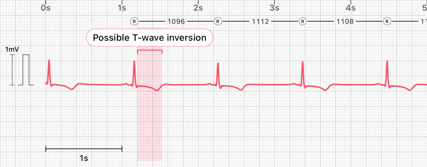
This is a real recording in lead I, the lead closest to what an Apple Watch records. ECG Tracker+ analyzed it with the same analysis it runs on your recordings.
Every QRS spike points up, as it normally does in lead I. After each one, the T wave dips below the baseline instead of rising above it. The app found an inverted T wave on all eight beats of the recording and marked a possible T-wave inversion, highlighting one beat as an example.
The cardiologists who read this person's full 12-lead ECG reported several other findings as well. Clinicians read the T wave together with everything else on the ECG and with the person's history, never on its own.
What can cause T-wave inversion
T-wave inversion is a sign, not a diagnosis. Possible causes include:2
- reduced blood flow to the heart muscle (ischemia) or an earlier heart attack
- a thickened heart wall, as in ventricular hypertrophy
- bundle branch block, which changes both the QRS and the T wave
- hypertrophic cardiomyopathy, a condition that thickens the heart muscle
- a blood clot in the lungs (pulmonary embolism)
- raised pressure inside the skull, for example after a brain injury
- a normal pattern in children, which persists into adulthood in some people
Only a clinician can tell which of these applies, usually with a 12-lead ECG and sometimes more tests.
Inverted T wave or upside-down recording?
If the Apple Watch is on a different wrist from the one selected in settings, the whole recording is flipped. Then the QRS spikes point down too, not only the T waves. The upside-down ECG guide explains how to fix the setting.
In a true T-wave inversion, the QRS still points up and only the T wave points down, as in the example. When the QRS of a recording points down, ECG Tracker+ is much more cautious, because the direction of the T wave is harder to judge. It then marks a possible T-wave inversion only when nearly every beat of a clear recording shows it.
How ECG Tracker+ decides
ECG Tracker+ checks the direction of the T wave on each beat it can read clearly. It marks a possible T-wave inversion only when a large share of those beats are inverted, so a single noisy beat does not trigger it. Small, flat T waves and noisy recordings can make the direction impossible to judge, and then no finding is shown.
What one lead cannot tell you
A 12-lead ECG shows the T wave from twelve angles. Clinicians look at which leads are affected and read the T wave together with the rest of the ECG.2 T-wave changes in the lateral leads, which include lead I, are common but nonspecific.3 One lead shows only one of those angles, which is why ECG Tracker+ calls it a possible T-wave inversion. It is a reason to show your recordings to a clinician, not a finding on its own.
Call emergency services for chest pain, pressure or tightness, fainting or severe shortness of breath, whatever any recording shows. If a possible T-wave inversion appears on your recordings and you have not discussed it before, bring them to a clinician.
Frequently asked questions
Is an inverted T wave dangerous?
It can be harmless or a sign of something important, depending on the cause. It needs a clinician to interpret, usually with a 12-lead ECG. If you have chest pain or other symptoms, seek care right away.
Can an Apple Watch detect T-wave inversion?
Apple's ECG app does not report the direction of the T wave. On a clear recording the T wave is often visible, and ECG Tracker+ marks a possible T-wave inversion for review.
Why does my whole Apple Watch ECG look upside down?
The most common reason is a wrist setting that does not match the wrist you wear the Watch on. That flips every wave, not only the T wave.
Does a normal T wave on my Watch rule out a heart problem?
No. A single lead cannot show every part of the heart, and Apple states that the ECG app cannot detect a heart attack.4